A red area on your skin is getting bigger, and you are trying to decide what it might be. The question lyme disease rash vs cellulitis: how to tell the…
A red area on your skin is getting bigger, and you are trying to decide what it might be. The question lyme disease rash vs cellulitis: how to tell the difference is one of the highest-stakes comparisons in bite and sting first aid, because the two conditions move on different clocks and need different urgency. This article gives you the triage framework clinicians use, so you know what to watch, what to record, and when waiting is not an option.
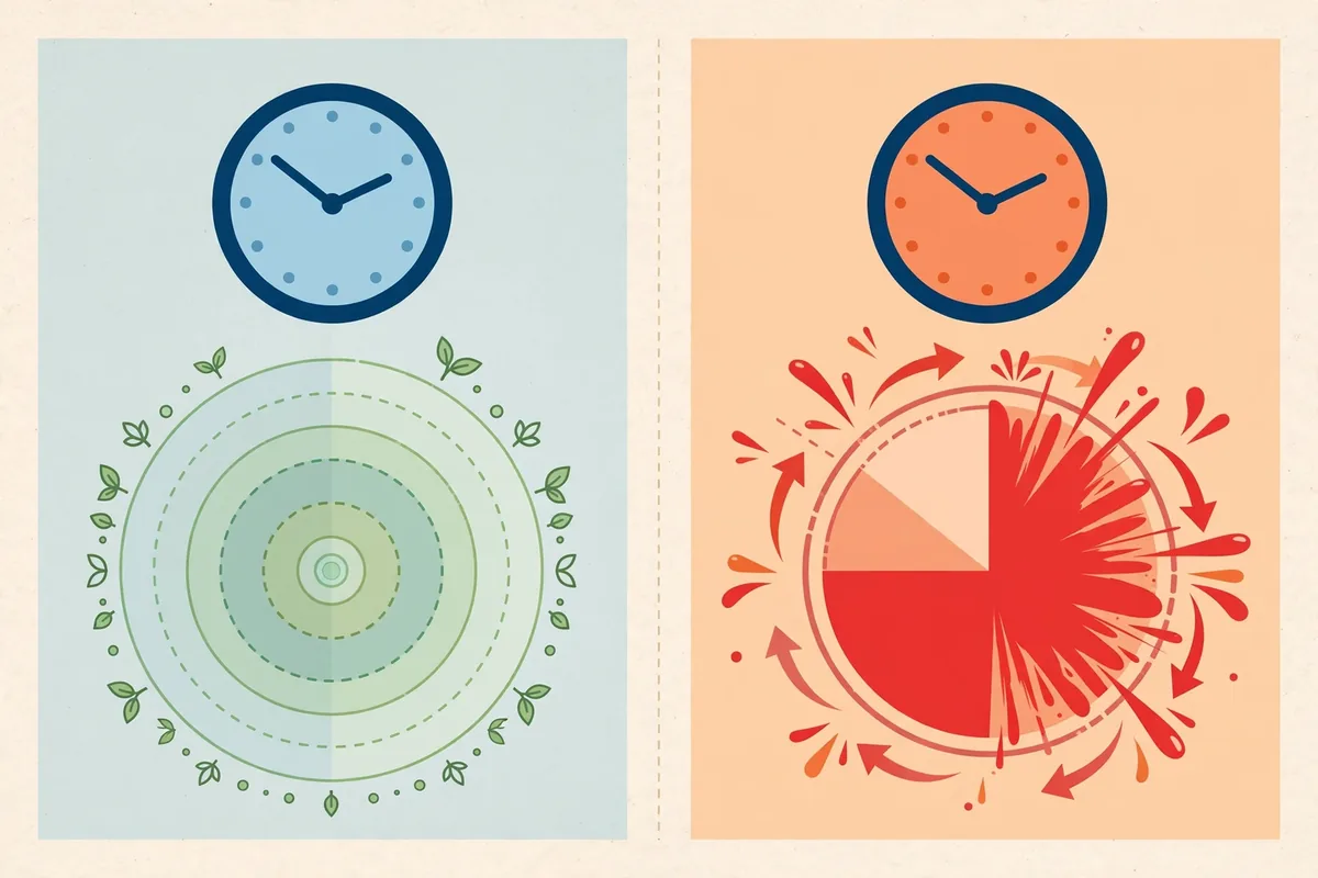
The direct answer: the Lyme disease rash, called erythema migrans, expands slowly over days to weeks, is usually flat, often feels warm but is rarely painful or itchy, and typically appears 3 to 30 days after a tick bite. Cellulitis, a bacterial skin infection, spreads over hours, is painful and tender to the touch, feels hot, and often comes with fever. Speed of spread is the single most useful fork in the road: growing over days points one way, spreading over hours points the other.
One boundary to set before anything else: this article cannot diagnose your rash, and you should not use it to diagnose yourself. Its job is to help you describe what you see accurately and decide how quickly you need a clinician. Any rapidly spreading, painful red area deserves same-day urgent care regardless of what you think caused it.
The Core Difference: Speed of Spread
Clinicians think about these two conditions in terms of time, and so should you.
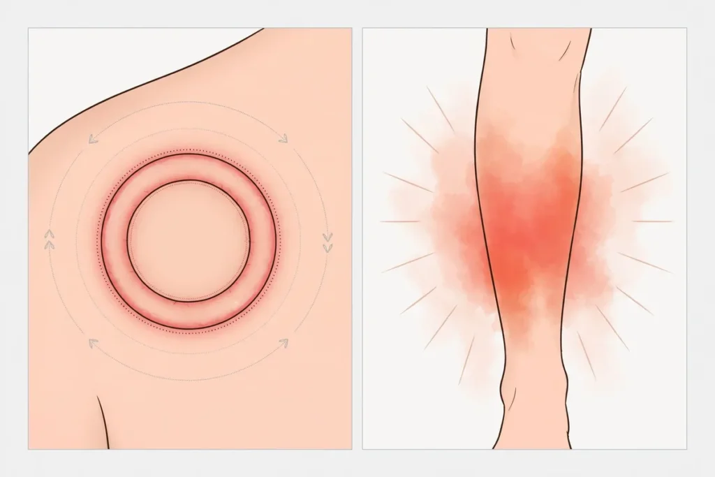
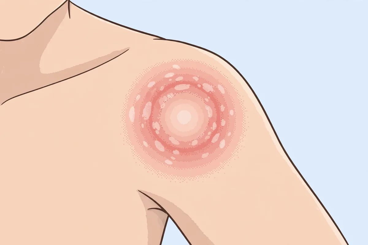
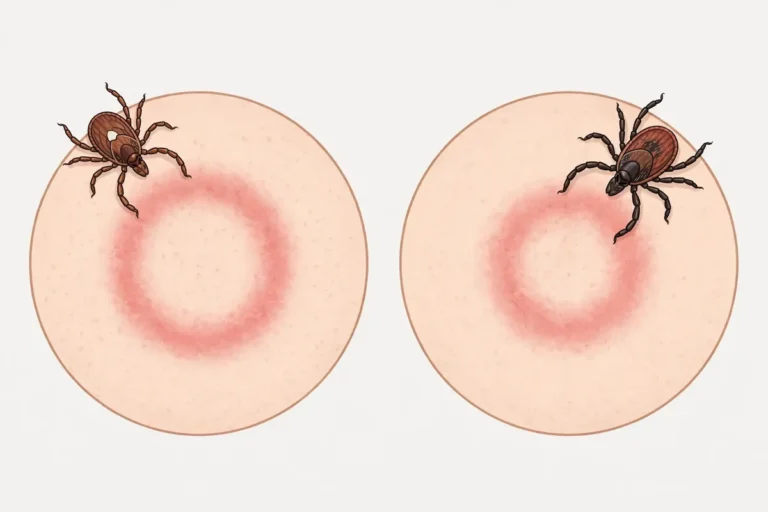
Erythema migrans, often abbreviated EM, is the rash of early Lyme disease. It begins at the site of a tick bite after a delay of 3 to 30 days, with about a week being typical, and then expands gradually over days. A single lesion can eventually reach 12 inches across or more. The expansion is steady and outward, sometimes with partial clearing in the center that creates the famous bull’s-eye appearance, though many EM rashes never form that pattern and stay uniformly pink or red. The key behavior: it keeps growing, day after day, rather than flaring and fading.
Cellulitis behaves nothing like that. It is a bacterial infection of the deeper skin layers, most often caused by streptococcal or staphylococcal bacteria entering through a break in the skin: a bite, a scratch, a crack between the toes. Once established, the red area expands over hours, and the time from first notice to a clearly spreading lesion is often measured in 6 to 36 hours. People frequently report that the redness visibly advanced between morning and evening. That pace is the red flag that separates it from EM.
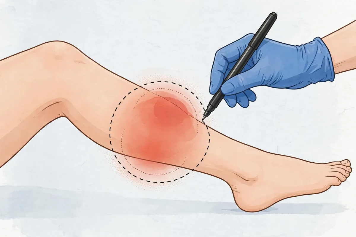
A practical way to use this: mark the border. Take a pen and lightly trace the edge of the red area, note the time, and check again in a few hours. If the redness has clearly crossed your line within hours, treat that as urgent. If the border creeps outward day by day over a week or more, note the pattern carefully and get prompt clinical evaluation, but the urgency profile is different. Either way, the tracing gives the clinician objective information, which is far more useful than a remembered impression.
What the Lyme Rash (Erythema Migrans) Looks Like
EM occurs in roughly 70 to 80 percent of people with Lyme disease, which means a meaningful minority never develop it, and its absence does not rule out infection. When it appears, it usually starts as a small red spot at the bite site and grows outward with a well-defined edge. The classic target or bull’s-eye look, a red center, a paler ring, then an outer red ring, is real but overrepresented in public imagination; many lesions are solid red or pink, some have a darker center, and a few blister slightly.
The surface is usually flat or only slightly raised and may feel warm to the touch. It is rarely itchy and almost never painful, which is precisely why people dismiss it. It can appear anywhere on the body, though ticks favor warm, hidden spots: the groin, armpits, backs of knees, waistline, scalp, and behind the ears. For surveillance purposes physicians look for a lesion at least 5 centimeters across, but you do not need to measure for a diagnosis; growth over days is the signal.
EM may be accompanied by flu-like symptoms: fatigue, headache, mild fever, muscle and joint aches, and swollen lymph nodes. Some people develop multiple smaller lesions as the infection spreads through the bloodstream, which is a sign the disease has disseminated and needs prompt treatment. Untreated, a single EM lesion can fade on its own within weeks, but the bacteria do not leave; the later stages of Lyme disease involve joints, nerves, and the heart. Fading is not healing.
One more caution about appearance: on brown or Black skin, EM can look like a bruise, a purplish or dusky patch, or an area only subtly darker than surrounding skin. Color matters less than behavior. A patch that is enlarging over days deserves evaluation whatever shade it is.
What Cellulitis Looks and Feels Like
Cellulitis announces itself. The affected area, most often one lower leg, but also arms or face, becomes red, swollen, warm, and painful or tender to touch. The borders are typically diffuse and poorly defined rather than sharp. The skin may look tight, shiny, or dimpled like orange peel as swelling builds, and blisters can form in more advanced cases. Red streaks extending outward from the area, called lymphangitis, signal the infection is traveling through the lymph system.
Pain is the great separator. EM is almost never painful; cellulitis hurts. Tenderness on light pressure, throbbing, and pain when moving the nearby joint are all typical. Systemic symptoms often follow: fever, chills, and a general feeling of being unwell commonly develop within a day or two of the skin changes, and sometimes fever precedes the visible redness. Swollen, tender lymph nodes near the area are common.
The entry point is often visible if you look: a healing insect bite, a scratch, a small cut, cracked skin between the toes from athlete’s foot. But sometimes no portal is obvious, and its absence does not rule out cellulitis. Cellulitis is almost never symmetrical; redness on both legs at once points toward other conditions like venous stasis and deserves its own evaluation.
Cellulitis can escalate. Warning signs of a deeper or more dangerous infection include severe pain out of proportion to what you see, numbness over the area, skin that turns dusky, purple, or black, blisters with bloody fluid, rapid spread despite treatment, and confusion, dizziness, or a fast heart rate. Those are emergency department signs, not wait-and-see signs.
Side-by-Side Comparison
Feature
Lyme rash (erythema migrans)
Cellulitis
Speed
Expands over days to weeks
Spreads over hours
Onset after trigger
3 to 30 days after tick bite, average about 7
Hours to a couple of days after skin break
Pain
Rarely painful, rarely itchy
Painful and tender to touch
Warmth
May feel warm
Noticeably hot
Borders
Well-defined, round or oval edge
Diffuse, poorly defined
Classic look
Expanding circle, sometimes bull’s-eye
Spreading redness, may streak outward
Fever
Mild fever possible with flu-like aches
Fever and chills common, can precede redness
Location
Anywhere; ticks favor hidden warm spots
Often one lower leg, arm, or face
Cause
Tick-borne bacterial infection
Bacteria entering through broken skin
Diagnosis
Clinical, based on appearance and history
Clinical, based on exam; sometimes blood work
No single row decides the matter. The pattern across rows is what clinicians weigh, and the two rows that carry the most weight are speed and pain: slow and painless leans toward EM; fast and painful leans toward cellulitis. When the picture is mixed, or when you cannot confidently place what you see, the safe move is clinical evaluation, not continued watching.
Tick Exposure History: What It Tells You
History is half the diagnosis, and your memory of the past month matters more than you might expect.
Ask yourself: have you been in woods, tall grass, or brushy areas in the past few weeks? Did you find a tick on yourself or a pet? Do you live in or did you travel through a region where Lyme disease occurs, which in the United States centers on the Northeast, upper Midwest, and parts of the West Coast, though the range has been expanding? A known tick bite, especially one where the tick was attached for a day or more, substantially raises the relevance of the EM side of this comparison.
But absence of a remembered bite proves nothing. Tick nymphs are the size of a poppy seed, their bites are painless, and most people with Lyme disease do not recall being bitten. So a blank tick history does not move you to the cellulitis column by itself; it just means the history is uninformative, and the rash’s behavior has to carry the decision.
Conversely, a clear alternative story strengthens the cellulitis side: a known cut, a scratched-open bug bite, or a skin crack that preceded the redness by a day or two fits the bacterial entry narrative. If you removed an attached tick recently, our guide on what to watch in the weeks after a tick bite lays out the monitoring timeline. And if you are unsure whether what you saw was a rash or just a local bite reaction, the tick bite rash versus local irritation comparison helps sort that earlier fork.
Why Lyme Blood Tests Can Be Negative Early
This section exists because the testing trap catches many people: a negative Lyme test in the first days of a rash does not rule out Lyme disease.
Standard Lyme testing is two-tiered: a screening test, usually an ELISA or EIA, followed by a confirmatory Western blot if the screen is positive or unclear. Both tests detect antibodies, your immune system’s response to the bacteria, not the bacteria itself. Antibodies take time to develop: IgM antibodies are generally detectable within about two weeks of infection, and IgG within about six weeks. Testing in the first days or weeks after a bite or rash onset is a well-documented cause of false-negative results.
For that reason, EM is a clinical diagnosis. Guidelines and clinical practice treat a characteristic expanding rash in the right context as sufficient basis for evaluation and treatment decisions, without waiting for blood work. Waiting weeks for antibodies to appear before acting would defeat the purpose of early treatment, which is to prevent dissemination.
Two related points deserve honesty. First, a positive test later does not distinguish active from past infection; antibodies can persist for years after successful treatment, so results always need clinical interpretation. Second, testing a tick itself is not a substitute for clinical judgment about the person. If a clinician evaluates your rash, bring the timeline: when you first noticed it, how fast it has grown, any tick exposure, and any systemic symptoms. That history is more diagnostically valuable than any single lab value in the early window.
When the Fork in the Road Cannot Wait
Some situations collapse the whole comparison into one instruction: get care now.
If a red area is spreading visibly over hours, is painful or tender, feels hot, and especially if fever, chills, or red streaks are present, that is same-day urgent care or emergency department territory. Do not wait to see whether it becomes EM-like; cellulitis can progress to bloodstream infection, and the treatment is antibiotics, which only a clinician can prescribe and calibrate.
If you have an expanding rash over days with a plausible tick exposure, that needs prompt clinical evaluation within days, not weeks. Early Lyme disease is highly treatable, and the benefit of evaluation is greatest before dissemination. This is not an emergency department visit at midnight, but it is not a next-month appointment either.
If the rash is expanding and you also develop facial droop, severe headache with neck stiffness, heart palpitations, or widespread joint swelling, seek urgent care; these can signal disseminated Lyme disease affecting nerves, heart, or joints.
And if you are ever unsure which column you are in, default to the faster one. It is far safer to have urgent care tell you a rash can wait than to wait on something that could not. For the broader picture of tick-borne risk after any bite, our Lyme disease after a tick bite guide covers the full timeline.
When to See a Doctor for an Expanding Rash
For any expanding red area after a bite or suspected bite, use these thresholds:
Same-day urgent care or ER: redness spreading over hours, significant pain or tenderness, heat, fever, chills, red streaks from the area, pus, blisters, dusky or blackened skin, or confusion and rapid heart rate
Prompt appointment within days: a rash expanding over days, especially with known or possible tick exposure in the past month, or with flu-like symptoms such as fatigue, headache, and body aches
Urgent re-evaluation: any diagnosed skin infection that worsens despite treatment, spreads rapidly, or develops new systemic symptoms
Routine follow-up: a small, stable bite reaction that is improving, with no spreading, no fever, and no tick exposure history, though photographing and dating it is still wise
Special cases: facial redness near the eye, redness in someone with diabetes, poor circulation, or a weakened immune system, all lower the threshold for urgent evaluation
Call 911 for difficulty breathing, throat or facial swelling, or dizziness after any bite or sting, which can indicate anaphylaxis rather than either condition discussed here. Our tick bite warning signs page collects the full set of thresholds in one place.
Frequently Asked Questions
Can a Lyme rash appear within hours of a tick bite?
A true erythema migrans does not. Redness within hours of a bite is a local hypersensitivity or irritation reaction, not EM, and surveillance definitions explicitly exclude annular redness appearing within hours of a tick bite from counting as EM. That early redness usually fades in a day or two. EM needs days to declare itself, which is why the monitoring window after a tick bite stretches to 30 days.
Does every Lyme rash look like a bull’s-eye?
No, and this misconception causes missed cases. Only a portion of EM lesions show the classic target pattern; many are uniformly pink or red, some have central darkening rather than clearing, and appearance varies with skin tone. The defining behavior is steady expansion over days, not any particular internal pattern. If you are waiting for a perfect bull’s-eye before seeking care, you may wait past the most treatable window.
Can cellulitis happen without a visible cut or bite?
Yes. The portal of entry is often microscopic: a tiny crack, dry skin fissure, or an unnoticed scratch. Athlete’s foot with fissures between the toes is one of the most common hidden entry points for lower-leg cellulitis. So the absence of an obvious wound does not argue against cellulitis when the speed, pain, and heat fit.
Should I draw a circle around the red area?
Yes, lightly tracing the border with a pen and noting the time is one of the most useful things you can do. It converts a vague impression of spreading into an objective measurement that any clinician can use. Take a dated photo as well. If the redness crosses your line within hours, that finding alone justifies urgent evaluation.
Can I use leftover antibiotics for a spreading red area?
No. Using someone else’s prescription, an old prescription, or an incomplete course is unsafe: the drug may be wrong for the organism, the dose may be wrong, and partial treatment can mask progression while the infection advances. Both cellulitis and Lyme disease need clinician-directed treatment. The urgency sections above tell you how fast to get that direction; the medicine cabinet is not a shortcut.
This article is for informational purposes only and isn’t medical advice. If you’re worried about a bite or sting, talk to a healthcare professional.
SJ
Dr. Sarah Jones, MD
MD — Medical Reviewer · Board-Certified Emergency Medicine Physician
Dr. Jones oversees StingHelp’s clinical verification process. Every guide is systematically evaluated against active Centers for Disease Control and Prevention (CDC), Environmental Protection Agency (EPA), and American Academy of Allergy, Asthma & Immunology (AAAAI) medical consensus standards to ensure symptom distinctions and care recommendations are clinically sound.
How this guide was prepared: Researched and structured by the StingHelp Editorial Team; verified against peer-reviewed dermatology literature; clinically reviewed November 2026.
RELATED CLINICAL GUIDES
More StingHelp guides
Explore physician-reviewed first-aid procedures, identification clues, and symptom comparisons for other common outdoor bites and stings.
One tick, six different clocks. Powassan virus can pass within 15 minutes while Lyme disease usually needs 36 to 48 hours — and official sources disagree on the hours in between. Every figure sourced, every conflict named.